Correctly label the following anatomical features of the neuroglia. … Correctly label the following parts of a chemical synapse. Drag each image below into the figure in order to correctly represent the sequence of events regarding the transmission at a cholinergic synapse. Step 3 has already been done for you.
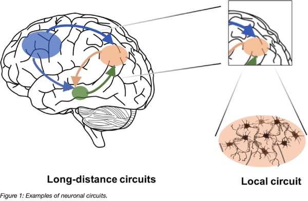
Dopamine Activates Astrocytes in Prefrontal Cortex via α1-Adrenergic Receptors | bioRxiv
Chemical synapses allow neurons to form circuits within the central nervous system. They are crucial to the biological computations that underlie perception and thought. They allow the nervous system to connect to and control other systems of the body. At a chemical synapse, one neuron releases neurotransmitter molecules into a small space (the

Source Image: tes.com
Download Image
Science Anatomy and Physiology Anatomy and Physiology questions and answers label the following parts of a chemical synapse Mitochondria Receptor Synaptic cleft Axon termina Synaptic vesicles Axon Neurotransmitter release This problem has been solved! You’ll get a detailed solution from a subject matter expert that helps you learn core concepts.

Source Image: quizlet.com
Download Image
Premium Vector | Synaptic cleft axon terminal science vector illustration graphic template The neuron or muscle cell receiving the information is called the postsynaptic neuron.On its surface, specialized proteins called receptors bind to the signaling molecule. For example, at the neuromuscular junction between the vagus nerve axon terminals and the heart, acetylcholine binds to muscarinic acetylcholine receptors.Between a spinal nerve axon terminal and the skeletal muscle it

Source Image: blog.addgene.org
Download Image
Correctly Label The Following Parts Of A Chemical Synapse.
The neuron or muscle cell receiving the information is called the postsynaptic neuron.On its surface, specialized proteins called receptors bind to the signaling molecule. For example, at the neuromuscular junction between the vagus nerve axon terminals and the heart, acetylcholine binds to muscarinic acetylcholine receptors.Between a spinal nerve axon terminal and the skeletal muscle it Resources. For the nervous system to function, neurons must be able to communicate with each other, and they do this through structures called synapses. At the synapse, the terminal of a presynaptic cell comes into close contact with the cell membrane of a postsynaptic neuron. Figure 8.1. The terminal of a presynaptic neuron comes into close
Using AAV for Neuronal Tracing
Start studying Synapse labeling. Learn vocabulary, terms, and more with flashcards, games, and other study tools. What Is a Neuron? Diagrams, Types, Function, and More

Source Image: healthline.com
Download Image
From Synapses To Switches: A Journey Through The Mystery Of Memory | IFLScience Start studying Synapse labeling. Learn vocabulary, terms, and more with flashcards, games, and other study tools.

Source Image: iflscience.com
Download Image
Dopamine Activates Astrocytes in Prefrontal Cortex via α1-Adrenergic Receptors | bioRxiv Correctly label the following anatomical features of the neuroglia. … Correctly label the following parts of a chemical synapse. Drag each image below into the figure in order to correctly represent the sequence of events regarding the transmission at a cholinergic synapse. Step 3 has already been done for you.

Source Image: biorxiv.org
Download Image
Premium Vector | Synaptic cleft axon terminal science vector illustration graphic template Science Anatomy and Physiology Anatomy and Physiology questions and answers label the following parts of a chemical synapse Mitochondria Receptor Synaptic cleft Axon termina Synaptic vesicles Axon Neurotransmitter release This problem has been solved! You’ll get a detailed solution from a subject matter expert that helps you learn core concepts.

Source Image: freepik.com
Download Image
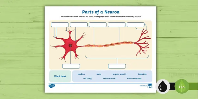
Parts of a Neuron Labelling Activity (teacher made) – Twinkl At a synapse, one neuron sends a message to a target neuron—another cell. Most synapses are chemical; these synapses communicate using chemical messengers. Other synapses are electrical; in these synapses, ions flow directly between cells. At a chemical synapse, an action potential triggers the presynaptic neuron to release neurotransmitters.

Source Image: twinkl.co.uk
Download Image
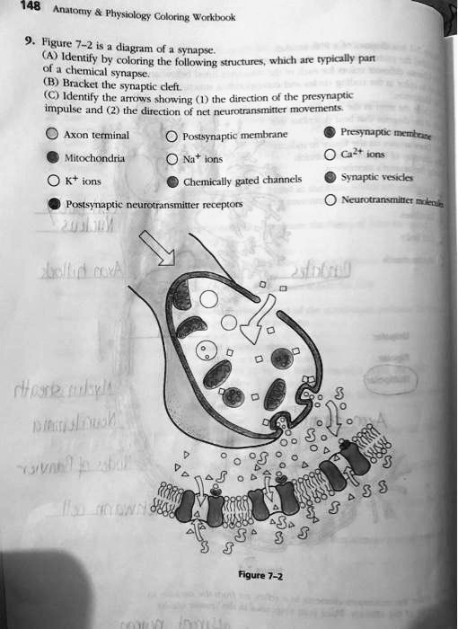
SOLVED: Please label the diagram of a synapse. Read instructions. (I’m not sure whether I’m right about the other colors, so you can label it too) 148 Anatomy Physiology Coloring Workbook 9. The neuron or muscle cell receiving the information is called the postsynaptic neuron.On its surface, specialized proteins called receptors bind to the signaling molecule. For example, at the neuromuscular junction between the vagus nerve axon terminals and the heart, acetylcholine binds to muscarinic acetylcholine receptors.Between a spinal nerve axon terminal and the skeletal muscle it

Source Image: numerade.com
Download Image
Neurobiological Mechanisms of Nicotine Reward and Aversion | Pharmacological Reviews Resources. For the nervous system to function, neurons must be able to communicate with each other, and they do this through structures called synapses. At the synapse, the terminal of a presynaptic cell comes into close contact with the cell membrane of a postsynaptic neuron. Figure 8.1. The terminal of a presynaptic neuron comes into close

Source Image: pharmrev.aspetjournals.org
Download Image
From Synapses To Switches: A Journey Through The Mystery Of Memory | IFLScience
Neurobiological Mechanisms of Nicotine Reward and Aversion | Pharmacological Reviews Chemical synapses allow neurons to form circuits within the central nervous system. They are crucial to the biological computations that underlie perception and thought. They allow the nervous system to connect to and control other systems of the body. At a chemical synapse, one neuron releases neurotransmitter molecules into a small space (the
Premium Vector | Synaptic cleft axon terminal science vector illustration graphic template SOLVED: Please label the diagram of a synapse. Read instructions. (I’m not sure whether I’m right about the other colors, so you can label it too) 148 Anatomy Physiology Coloring Workbook 9. At a synapse, one neuron sends a message to a target neuron—another cell. Most synapses are chemical; these synapses communicate using chemical messengers. Other synapses are electrical; in these synapses, ions flow directly between cells. At a chemical synapse, an action potential triggers the presynaptic neuron to release neurotransmitters.